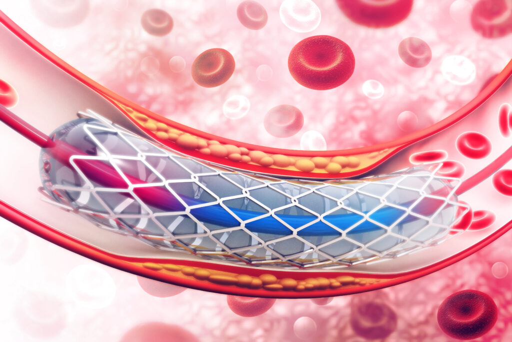
A venogram is imaging of a vein obtained with a small amount of contrast dye and/or carbon dioxide. This imaging can be both diagnostic and/or for treatment. This is done by making a small nick in the skin at the access site, usually the groin. A thin catheter is then placed into the vein for imaging and potential treatment. A special ultrasound catheter placed inside the blood vessel gives the most accurate information of the degree of compression and vessel measurements for precise treatment. This imaging allows our Vascular Interventional Radiologists to see if there is compression of the Iliac vein by the Iliac artery. If there is significant compression, a stent will then be permanently placed into the vein. A balloon, called a venoplasty, is then placed into the vein temporarily inflated to enlarge the vein and ensure proper stent placement. This can help with diagnosing and treating May-Thurner.
A stent is a metal mesh tube that is inserted permanently into an artery to help keep the vessel open. The stent is placed using the imaging from an angiogram and is placed by a Vascular Interventional Radiologist. The placement of a stent is often used in conjunction with an angioplasty. The procedural steps are very similar, but instead of a balloon, the stent is what holds the artery open. This ensures consistent blood flow after the procedure is complete. The placement of a stent is very precise and choosing an expert in this field is important, much like our Vascular Interventional Radiologist at Peripheral Vascular Partners.

While each therapy has its own set of advantages and applications, a venogram is best suited for patients suspected from clinical history of symptoms to diagnose whether or not May Thurner is the cause and allows for simultaneous treatment.
A Venogram can diagnose and help address MTS before it forms a blockage large enough to cause deep vein thrombosis. A Venogram has a much faster recovery time than surgical alternatives. It is also a same day procedure, so you will be home and recovering in the comfort of your own residence.
Venograms are minimally-invasive, and therefore do not result in many of the issues often associated with major surgery. This means they do not utilize a large incision, which can cause significant scarring, nor do they use general anesthesia. The patient is comfortably sedated, but remains conscious but sleepy during the duration of the treatment.
The next step is to choose a facility to perform your venogram. In doing so, make sure you select a team of well trained specialists, who utilize the best available medical technology to perform their procedures.
If you want to ensure you receive the best quality procedure and safest experience for your venogram, you’ll want to choose Peripheral Vascular Partners.
At Peripheral Vascular Partners, we understand that MTS and deep vein thrombosis are a serious matter. We make it our mission to walk you step-by-step through your procedure, ensuring you are comfortable during each phase of your treatment. Our specialists are experts in vascular treatments, and treat each patient with the same level of focus and care.
If you are looking for a less invasive procedure a venogram may be a great option for relief from MTS without the risk and discomfort of invasive procedures. We are here to answer any questions you have about the procedures involved with our MTS treatments. Simply schedule a consultation today!